
Services
Removal of facial and oral neoplasms
Surgical removal of benign formations under operating-room conditions.
from 20 000 ₸ Per lesion; histology is charged separately
- MD surgeons
- 3D planning
- International standard
WHAT PATIENTS COME WITH
Common concerns
IN DETAIL
About the procedure in detail
Why removal ends in the laboratory
Why removal ends in the laboratory
The growth is removed whole and sent for histological examination. The laboratory examines the cells under a microscope and gives an answer as to what it was. There is no other way to know for certain: nobody can tell a dangerous growth from a harmless one by its appearance.
That is why we do not burn off or shave off growths on the face by methods that leave nothing to examine. If the doctor sees signs calling for a different scope of care, he says so at the examination and refers the patient to the relevant specialist instead of removing the growth "just in case".
Which growths people come with
Which growths people come with
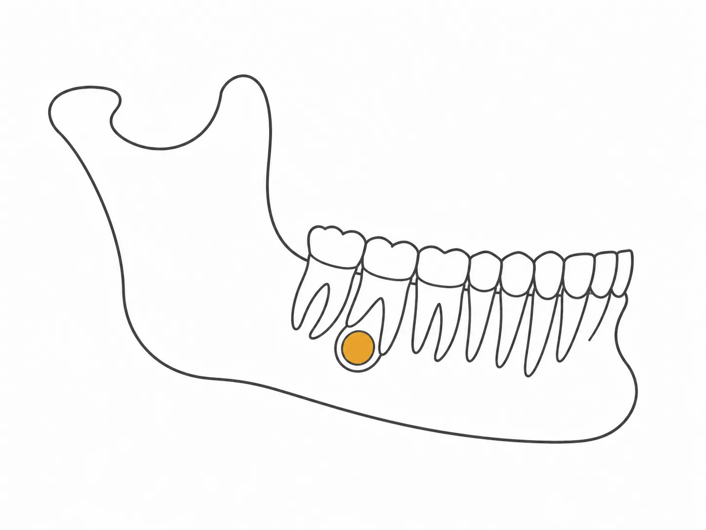
Moles, papillomas, cysts and other formations of the facial skin - including those traumatised by a razor, by clothing or by glasses. Lumps on the gum, the cheek, the tongue and the mucosa that interfere with chewing or are caught by a denture.
If the growth lies deeper and its borders are not visible on examination, the scope of the procedure is determined from the imaging.
How the removal goes
How the removal goes
The surgeon examines the growth and asks how long ago it appeared and what has happened to it since. Where necessary he orders imaging or an additional examination to establish its depth and borders.
The removal itself is performed under local or general anaesthesia - depending on the scope. The direction of the incision is chosen along the skin lines so that the trace is as inconspicuous as possible. The removed tissue is sent to the laboratory.
The doctor reports the result of the examination, explains what it means and says whether further monitoring is needed. Histology is not sent as a file without comment.
What is discussed before the removal
What is discussed before the removal
Removing a growth is a surgical operation, even when the growth is small. A trace at the site of the incision always remains: the surgeon's task is to make it as inconspicuous as possible, not to promise its absence. How visible it will be depends on the size, the location and the healing.
In the first days swelling and firmness in the treated area are possible. If the removal is incomplete, the growth may appear again. If the histology shows that a different scope of care is needed, the doctor will refer the patient to the relevant specialist.
The result is always individual and depends on the diagnosis, the preparation, the scope of treatment and the recovery.
FIND YOUR TREATMENT
Not sure what suits you?
Answer a few questions and the doctor will advise which direction suits you best.
HOW TREATMENT WORKS
How treatment works
-
01
Consultation
The doctor examines the growth and determines the removal approach
-
02
Diagnostics
Additional tests are ordered before the procedure if needed
-
03
Surgery
The growth is excised under operating-room conditions
-
04
Histology
The removed tissue is sent for analysis
-
05
Follow-up
The doctor monitors healing and communicates the histology result
BENEFITS
Benefits
Histology testing
A minimal trace
A licensed operating room
Follow-up after surgery
THE TEAM
Specialists


REVIEWS
Patient reviews
A mole on my cheek started changing colour and growing slightly; my dermatologist recommended removing it and sending it for histology. The clinic did everything quickly - examination, removal, and sending the tissue for analysis. The histology result was reassuring, and the scar is barely visible after a month.
A papilloma on my chin kept catching on my clothes or a towel, which was unpleasant. I went to the clinic, the doctor examined it and suggested surgical removal the same day. The procedure took very little time, and almost no trace remains. I regret putting up with it for so long instead of coming in sooner.
I noticed a small lump in my mouth, got worried, and went straight to a consultation. The doctor examined it, ordered diagnostics, and explained that removal under operating-room conditions was best for precision. Everything went smoothly, the tissue was sent for histology, and a week later I got the result - my worries turned out to be unfounded.
QUESTIONS
Frequently asked questions
Is removing a growth painful?
The procedure is performed under local or general anaesthesia depending on scope - the patient feels no pain during removal.
Is histology mandatory?
Yes, removed tissue is sent for analysis - this is standard practice for patient safety.
Will there be a scar?
The trace depends on the growth's size and location; the doctor explains what to expect beforehand.
How long does recovery take?
Healing usually takes from a few days to a couple of weeks, depending on the scope of the procedure.
MORE SERVICES
You may also be interested in


CONSULTATION
Book a Consultation
Free specialist advice